38 onion cells under microscope with labels
Leaf Cell Under Microscope Labeled - rohrreinigung-sued.de 1 day ago · leaf cell under microscope labeled. Leaf Cell Under Microscope Labeled. w7m, ur3, knj4, ... Natural Sciences Grade 9 - Grade 7-9 Workbooks The onion cells have a thick cell wall and a cell membrane. The animal cells only have a cell membrane. The onion cells have a regular shape whereas the cheek cells have a irregular shape and seem more flimsy. In the onion cells they might notice a large vacuole which might not be as visible in the cheek cells. Cheek cells do not have vacuoles ...
The Biology Project The Biology Project, an interactive online resource for learning biology developed at The University of Arizona. The Biology Project is fun, richly illustrated, and tested on 1000s of students.

Onion cells under microscope with labels
Plant Cell Under Microscope Labeled 40X : Young Root 2 Of Broad Bean ... Cells and viewing them under the microscope. A small square of a red onion skin (membrane) was observed under a microscope at high power (x40) magnification. (iv) describe how you applied the stain. They must draw and label the nucleus, cell membrane set up your microscope, place the onion root slide on the stage and focus on low (40x) power. Observing Onion Cells Under The Microscope Afterwards, carefully mount the prepared and stained onion cell slide onto the microscope stage. Make sure that the cover slip is perfectly aligned with the microscope slide, and that any excess stain has been wiped off. Secure the slide on the stage using the stage clips. PDF Onion Cells - Investigation - Exploring Nature 5. Observe the onion tissue under the microscope at 4x, 10x and 40x with lots of light (open diaphragm). Then slowly close the diaphragm while observing the image to find the best light for seeing cellular details. 6. Draw a section of onion skin cells at 10x magnification. Then switch to 40x and draw one cell and label it. Questions: 1.
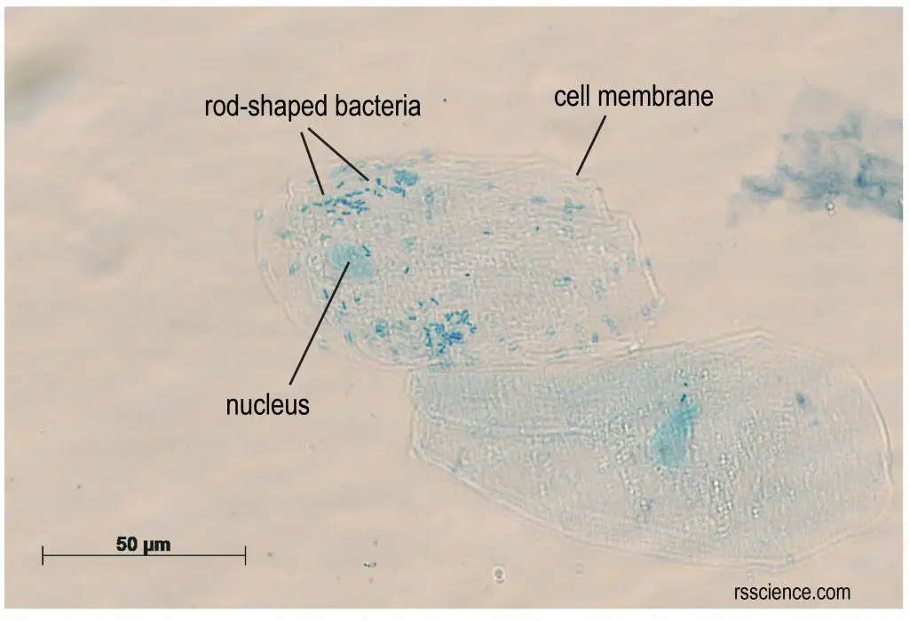
Onion cells under microscope with labels. Plant tissue under a microscope - xylem and phloem - Rs' Science The highly active mitosis area is highlighted with a red dash line. Within that area, you can easily find cells undergoing different phases of mitosis, prophase , metaphase , anaphase, and telophase. (Modified from the guidebook of Rs' Science - 25 Microscope Prepared Slide Set) The Stem - Xylem and Phloem PDF Onion Cell Lab - somewaresinmaine.com Research Biology Onion Cell Lab page 1 of 3 Onion Cell Lab After you have completed the rest of this lab come back to this cover page DRAW & LABEL AN ONION CELL WITH ALL THE PARTS / ORGANELLES YOU OBSERVE UNDER 40X. Purpose: To observe and identify major plant cell structures and to relate the structure of the cell to its function. Materials: 1 ... Animal Cell Under Light Microscope Labelled : Draw and label the ... Onion cell diagram labeled structure of animal cell and plant cell under microscope. An organelle found in large numbers in most cells, in which the biochemical processes of respiration and energy production occur. Under a light microscope, the cell membrane, nucleus and cytoplasm of a cheek cell (animal cell) can be observed. Onion Epidermal Cell Labeled Diagram - schematron.org Draw a labelled diagram of an onion epidermal cell seen under the microscope. ( 4 marks) e The onion epidermal cells are not green in colour because they lack. The epidermal cells of onions provide a protective layer against viruses and fungi that may harm the sensitive tissues.
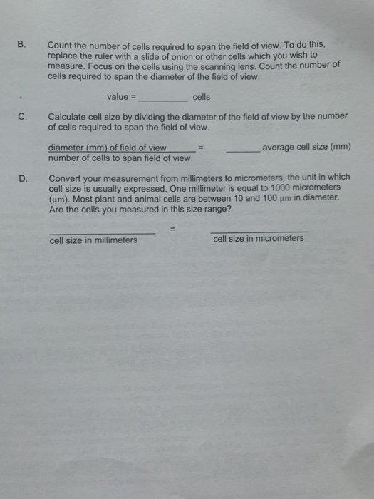
DOC Plant and Animal Cells Microscope Lab - Hillsboro City Schools Make a drawing of one onion cell, labeling all of its parts as you observe them. (At minimum you should observe the nucleus, cell wall, and cytoplasm.) Cheek cells 1. To view cheek cells, gently scrape the inside lining of your cheek with a toothpick. DO NOT GOUGE THE INSIDE OF YOUR CHEEK! (We will observe blood cells in a future lab!!) 2. Onion Cells Microscope Stock Photos and Images - Alamy Onion cells under the microscope. Garden onion, Bulb Onion, Common Onion (Allium cepa), cell tissue of a garden onion with dyed chromosomes, light microscopy, x 120, Germany. Onion Cells under the Microscope. Onion skin cells under the microscope, horizontal field of view is about 0.61 mm. Detailed view of the cells of a red onion as seen ... Plant Cell Under Microscope 40X Labeled : 1 - Chloroplast and cell wall ... The different images below were taken with two different types of microscopes. 1.can only turn fine adjustment 2.draw one row of cells across the middle 3.label the chloroplasts and cell wall. When using the microscope always start by focusing under low power and working your way up to high power. Microscopy, size and magnification - Microscopy, size and ... - BBC Place cells on a microscope slide. Add a drop of water or iodine (a chemical stain). Lower a coverslip onto the onion cells using forceps or a mounted needle. This needs to be done gently to...
Onion Epidermis - kuensting.org Onion epidermal cells, iodine stain, 400X. The nucleus of an onion epidermal cell, 1000X magnification. ... rhoeo discolor leaf under microscope labeled - Erotske priče … In contrast, the light has to pass through the specimen to form the image under a compound microscope. Cutter 6. No need to register, buy now! Zebrina. Take an onion bulb/ rhoeo leaf, with the help of forceps pull a thin transparent peel. In Microscope Lab II, we look at the __ of a leaf of the Rhoeo discolor plant to see representative plant cells. Also note the time taken to plasmolysis ... Animal Cell Diagram Under Microscope Labeled Animal Cell Diagram Under Microscope. Function cell does in the body dictate the change and adaptation done by cell. When observing onion cells, there is the Cell Surface Membrane which is present in all living cells. We all keep in mind that the human body is quite intricate and a method I discovered to are aware of it is via the manner of ... DOC The Onion Cell Lab - chsd.us Onion tissue provides excellent cells to study under the microscope. The main cell structures are easy to see when viewed with the microscope at medium power. For example, you will observe a large circular . nucleus. in each cell, which contains the genetic material for the cell. In each nucleus, are round bodies called . nucleoli

Plant Cells Under Microscope Labeled - Micropedia
Lennox Educational 01: To use a light microscope; 02: To obtain a good specimen of plant tissue for viewing under the microscope (onion cells) 03: To obtain a good specimen of animal tissue for viewing under the microscope (cheek cells) 04: To investigate the digestion of starch by amylase; 05: To investigate the effect of exercise on heart rate
Light Microscope Onion Cell Labeled - Micropedia
Oxford Cambridge and RSA Friday 16 October 2020 – Morning 1 (a) A student was observing onion epithelial cells using a light microscope. They photographed these cells and the image obtained is shown in Fig. 1.1. The student then made a drawing of a few cells from this image. The drawing is shown in Fig. 1.2. Fig. 1.1 cytoplasm cell wall large permanent vacuole ribosome Fig. 1.2
Animal Cell Under Microscope 1000X : How These 26 Things Look Like Under The Microscope With ...
Under the Micrsocope: Onion Cell (100x - 400x) - YouTube In this "experiment" we will see onion cells under the microscope.For the experiment you will only need onion, dropper and the microscope (container and tool...
Onion Root Images
Leaf Cell Under Microscope Labeled 1 day ago · A student draws a leaf and labels it ½ X. Leaf Cell Under Microscope Labeled - Micropedia. The most common inquiries are how you may differentiate the duodenum, jejunum, and ileum histology slide. Diagrams Of Monocot Leaf Under The Microscope plant cells under microscope stock images download 633.

General Biology Microscopic Specimen Images & Photographs
The Cell - ScienceQuiz.net The diagram shows a group of onion cells. The parts labelled A, B and C respectively are ... The diagram shows a plant cell as seen under a microscope. Two of the ...
Post a Comment for "38 onion cells under microscope with labels"